Enhancer chromosomal-mediated chromosome hijacking and increasing deposition of acetylated histone H3 lysine 27 (H3K27ac) can support oncogenic expression in cancer cells. However, it is not clear how the chromatin of enhancer-promoter interactions is affected by these events.
In a new study, researchers at the EPFL and UNIL have used a new algorithmic approach to study how cancer cells can restructure their DNA’s 3D structure in order to increase the activity of “oncogenes” genes that promote cancer.
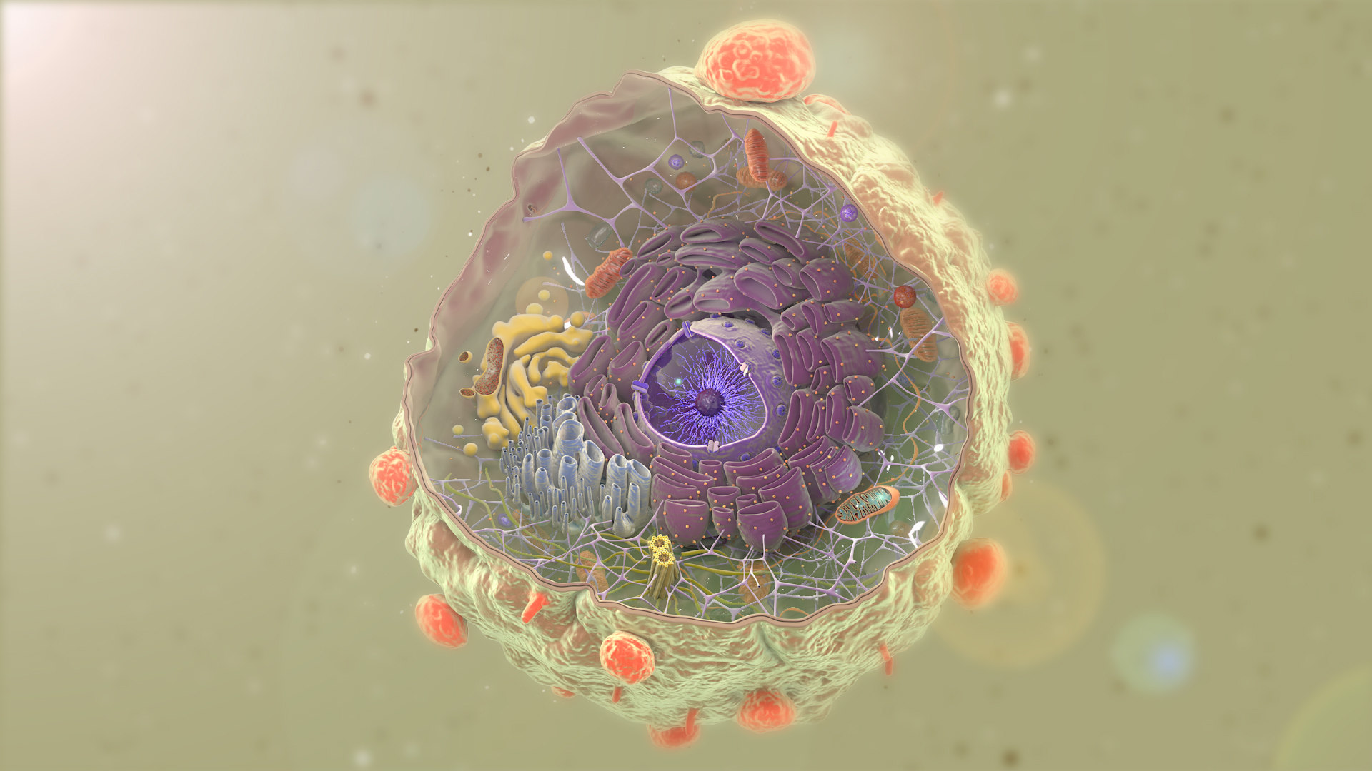
Scientists focused primarily on chromosomes and their organisation in the narrow space of the cell nucleus.
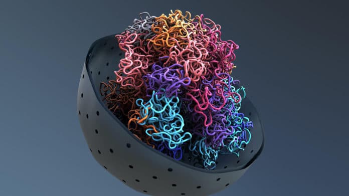
Each cell in our bodies has about two metres of DNA so that we have evolved to store it properly, understandably. This mechanism involves winding DNA around specific proteins known as histones, which form a chromatin-protein complex.
Several chromatins form the chromosome structure. Researchers examined how the changes in explicit epigenetic marks modify chromosome structures and the expression of genes known as oncogenes that promote tumour growth.
Giovanni Ciriello’s UNIL team has developed a new algorithmic approach to track the genomic regions in the nucleus, called Calder (after the American sculptor, Alexander Calder).
“We used Calder in more than 100 samples to compare the spatial organisation of the genome. But it is not static, and like the mobile sculptures of Alexander Calder, it can rearrange its pieces.”
Using Calder, scientists determined which regions of chromatin were moved by changing epigenetic marks from one area of the nucleus to another. These changes have been observed in normal lymphoma cells and B-cells.
They found that some epigenetic changes, particularly in lymphoma cells, lead to repositioned areas of chromatin in different areas of the nucleus and foster local interactions that overactivate oncogene expression.
It was also discovered that when two fragments of different chromosomes are broken and exchanged, they assume a 3D structure that is different from normal copies. Importantly, these changes in 3D structure are in line with different epigenetic marks and induce high levels of genes supporting the expansion of tumour cells.
Elisa Oricchio (EPFL) said, “We usually see our DNA as a long, linear molecule, and only recently we began to understand the way in which their 3D organisation is altered in cancer cells. The spatial organisation of DNA in the nucleus provides a new lens to understand how tumour cells originate and how epigenetic mark therapeutic modulation can prevent the progression of tumours.”
_____________________________________________________________
Cancer | Don’t forget to follow us on Twitter @njtimesofficial. To get latest updates